Insulin, 1955: A Physician’s Read on the First Protein Sequence
By Andres Zuleta, MD · Prefer the patient version? Read it on the ThriveMed blog.
Insulin was in clinical use for 33 years before anyone could write down its structure. One of the first children treated, Elizabeth Hughes, began injections on August 17, 1922 and lived 58 years on insulin (University of Toronto, Hughes Papers); by one estimate she received about 42,000 doses. Frederick Sanger's Biochemical Journal papers of 1951, 1953 and 1955 closed that gap, and they deserve a careful read because they settled a question we now take for granted: whether a protein has one defined sequence at all.
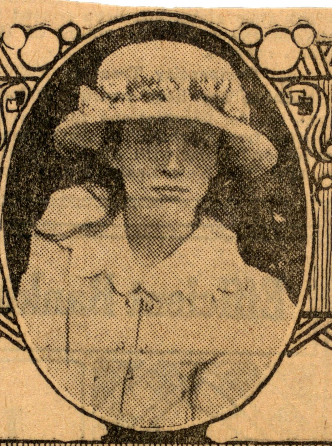
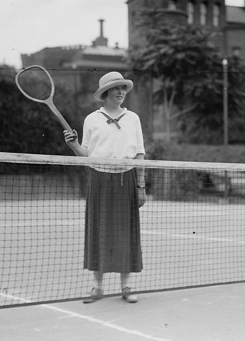
Left: Elizabeth Hughes as printed in the Toronto Daily Star on August 17, 1922, the day of her first injection (public domain, published 1922; scan courtesy of the Thomas Fisher Rare Book Library, University of Toronto, insulin:C10033; the date the photograph was taken is not recorded). Right: Elizabeth in 1924 (National Photo Company, Library of Congress, public domain). Her own chart records a 9 pound gain by September 3, 1922.
Why this belongs in a peptide series: insulin is a peptide hormone, two chains totaling 51 residues. It sits at the conventional boundary between peptides and proteins (US regulation, 21 CFR 600.3(h)(6), counts chains associated in nature together and calls anything over 40 residues a protein, which is why insulin is licensed as a biologic). Sanger's work made it the first peptide, and the first protein, with a known sequence.
The question in 1943
Sanger's Nobel lecture (Dec 11, 1958) describes the field he entered: amino acid composition could be measured, but "practically nothing" was known about order. The competing models were Bergmann and Niemann's periodicity hypothesis and the view that a purified protein was a statistical mixture of similar chains. Insulin was a practical choice: Chibnall's group had an accurate analysis, it lacked tryptophan and methionine, and it had a high content of free alpha-amino groups, suggesting short chains. For most of the project its molecular weight was believed to be 12,000; the true monomer is about 6,000.
The method
The 1945 paper introduced 1-fluoro-2,4-dinitrobenzene (FDNB) to label free amino groups; after acid hydrolysis the N-terminal residue is recovered as a yellow DNP derivative. Performic acid oxidation split the disulfides and separated a glycyl (A) and a phenylalanyl (B) chain. Partial acid and enzymatic hydrolysis then produced overlapping peptides, which were fractionated by paper chromatography and ionophoresis and assembled into a unique sequence. The B chain (30 residues) appeared in September 1951 (Sanger and Tuppy), the A chain (21 residues) in February 1953 (Sanger and Thompson). The disulfide assignment (Ryle, Sanger, Smith and Kitai, August 1955) was the hardest step, because disulfide interchange during acid hydrolysis scrambled the bridges until it was suppressed with thiol reagents. The result: A7 to B7 and A20 to B19 interchain bridges plus the A6 to A11 intrachain loop.
The conclusion that mattered
"Proteins are definite chemical substances possessing a unique structure in which each position in the chain is occupied by one and only one amino acid residue." The sequence showed no periodicity, "a random order, but nevertheless a unique and most significant order." That is the conceptual hinge between protein chemistry and the genetic code. It is also why species comparisons became informative: cattle, pig, sheep, horse and whale insulins share the B chain and differ only at A8 to A10, inside the intrachain disulfide loop (Brown, Sanger and Kitai 1955; Nobel lecture). Human insulin (Nicol and Smith, Nature, August 1960) differs from porcine insulin only at B30 (threonine vs alanine) and from bovine at A8, A10 and B30, which I confirmed today against UniProt entries P01308, P01315 and P01317.
What the sequence could not tell us
- Conformation. The three-dimensional structure required X-ray crystallography (Adams, Hodgkin and colleagues, Nature 1969).
- Biosynthesis. The two-chain molecule is processed from a single-chain precursor, proinsulin (Steiner and colleagues, Science, August 1967). The excised connecting peptide, C-peptide, is cosecreted with insulin.
- Throughput. About twelve years for 51 residues. Automation came with Edman and Begg's sequenator (Eur J Biochem 1967), and Sanger's later dideoxy method (PNAS 1977) moved the field to reading genes rather than proteins.
The clinical line from 1955
- Single-residue disease. Ingram's 1956 and 1957 Nature papers localized the sickle hemoglobin defect to one amino acid using peptide fingerprinting built on Sanger's approach.
- Rational analogs. Insulin lispro reverses B28 proline and B29 lysine and is labeled rapid-acting (FDA label; approved June 14, 1996, BLA 020563). Every analog since is a sequence edit.
- C-peptide in practice. The ADA Standards of Care 2026 (section 2, Figure 2.1) use C-peptide with a concurrent glucose to classify diabetes in insulin-treated adults: 600 pmol/L or more supports type 2, very low values (under 80 pmol/L) indicate severe insulin deficiency, intermediate values need context; avoid testing within 2 weeks of a hyperglycemic emergency.
- Scale. UniProtKB/Swiss-Prot lists 575,748 reviewed sequences (20,431 human) and 149,430,635 unreviewed sequences in release 2026_03 (September 2, 2026).
For patients
What I share in clinic: every legitimate peptide medicine has a defined, published sequence, so "what exactly is in it?" is a fair first question for anything sold as a peptide. And if you are treated with insulin, C-peptide is a simple test that can clarify how much insulin your pancreas still makes.
For the patient-friendly version, read my post on the ThriveMed blog: What Was in the Medicine That Saved Elizabeth Hughes? The First Protein Ever Read. Join Thrive Nation: https://thrivemed.ai/programs/human-health-os?utm_source=drzuleta&utm_medium=blog&utm_campaign=peptide_breakthroughs_ep4&utm_content=cta#thrive-nation
The Peptide Breakthroughs: Previous, Episode 3: Oxytocin, 1953: the first synthetic peptide hormone · Next, Episode 5 (link when live).
Want a proactive plan for your own health? Join Thrive Nation.
Sources: Toronto Daily Star, Aug 17, 1922 (U of T insulin:C10033); 21 CFR 600.3(h)(6); Biochem J 1945;39:507-515; 1951;49:463-481 and 481-490; 1953;53:353-366 and 366-374; 1955;60:541-556 and 556-565; Sanger F, Nobel lecture, Dec 11, 1958; Nature 1956;178:792-794; 1957;180:326-328; 1960;187:483-485; Science 1967;157:697-700; Eur J Biochem 1967;1:80-91; Nature 1969;224:491-495; PNAS 1977;74:5463-5467; FDA Humalog label (BLA 020563); Diabetes Care 2026;49(Suppl 1):S27-S49; UniProt release 2026_03; University of Toronto, Elizabeth Hughes Papers. Educational only, not medical advice.